Innovative software improves accuracy for liver surgery
A new imaging and tracking surgical software developed by a Washington University physician aids complicated liver surgeries

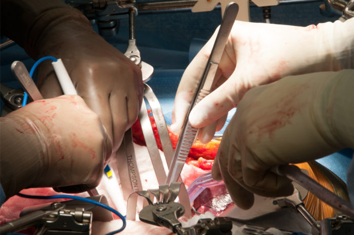
During liver surgery, William Chapman, MD, tests the accuracy of a 3D image-guidance system.
Liver resection – surgically removing all or part of the liver – is becoming more common as a treatment for liver cancer and disease. A Washington University physician has co-founded an FDA-approved image planning and operation guidance software to help with this form of surgery. The software generates a 3-D image from medical scanning technologies such as CT and MRI. It then accurately calculates the planned volume of liver to be removed and the volume that will be left in place. This helps liver surgeons plan for what are often complicated liver surgeries.
The technology, developed by Pathfinder Therapeutics, Inc., consists of an optical tracking system, tracked surgical instruments, and the image-guided surgery software that provides 3-D visualization of anatomic structures and real-time guidance of therapeutic delivery tools. William Chapman, MD, chief of the Division of General Surgery and chief of the Section of Transplantation at Washington University, is one of the founders of Pathfinder and began developing the technology 14 years ago.
Streamlining surgery
During surgery, the system tracks operative instruments using infrared emitters to within 1/10 of a millimeter in a standard operating room.
“When planning, I can see where I want to make the resection,” says Chapman. “But once I start the surgery, I can now correlate my pre-operative images with the real-time surgical location. I can register my position and track where I am inside the liver while making the resection.”
Using the 3-D pre-operative planning images, Pathfinder allows the surgeon to plan the resection and also measures the volume of liver removed, the volume left behind and the percentage of the remnant liver.
“That’s the key,” says Chapman. “When removing a tumor, it’s important to know what part of the liver is still functional and how much of the liver can be left without risk for reoccurrence in the patient. The pre-operative planning gives physicians assurance that the remnant liver is functional and adequate for the patient.”
Chapman says that his team still uses ultrasound, CT scanning and other imaging. “This is just another way to provide reassurance to surgeons as well as save time and provide a more streamlined way of surgical planning.”


