Clarifying connections between glaucoma, cataracts and oxygen levels
New findings show that elevated levels of oxygen in the eye appear to contribute to the development of cataracts and glaucoma

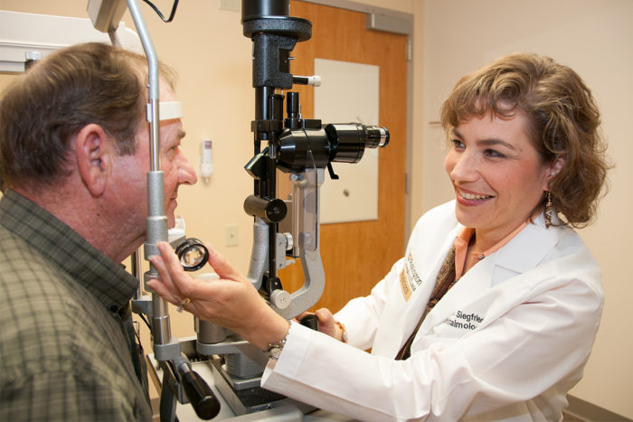
Glaucoma specialist Carla Siegfried, MD (right), examines Jerry Rann at the Washington University Eye Center.
Cataract surgery is so common in the US that it is the most expensive item in the Medicare ophthalmology budget. Meanwhile, glaucoma takes a disproportionate toll on African Americans, who are six times more likely than Caucasians to develop the disorder and 16 times more likely to go blind from it.
To learn how oxygen fits in, a research team, led by David Beebe, PhD, Ying-Bo Shui, MD, PhD, Carla Siegfried, MD, and collaborator Nancy Holekamp, MD, has measured oxygen levels in the eyes of patients having glaucoma surgery, cataract surgery and retina surgery. During operations, they insert probes to assess oxygen levels at several locations in the eye.
The retina procedures, which involve the removal of the vitreous gel that fills the eye, are important in these studies because it is nearly certain that these patients will develop a nuclear cataract (that affects the center of the eye’s lens) within two years following surgery.
Toxic levels of oxygen

In studies conducted over the last several years, Beebe has found that immediately after retina surgery, oxygen levels in the eye are about eight times higher than normal. This is because during a vitrectomy, where the vitreous gel is removed during the surgery, the fluid infused into the eye to replace it has high oxygen. Levels of oxygen near the lens eventually return to about one and one-half times normal, but by then, the oxygen already may have done significant damage.
“It may be possible to design interventions to protect the lens in people who have had a vitrectomy or whose vitreous is degenerating as a part of normal aging,” Beebe said.
Shui is now leading a research effort to do just that, and his experiments have found that it’s possible to restore the structure of the vitreous gel in the test tube and in animals.
During glaucoma surgery the researchers concentrated on oxygen measurements taken in the eye’s anterior chamber angle, where the cornea meets the iris. That region is particularly important in glaucoma because it is where fluid drains from the eye. If the fluid can’t drain properly, pressure builds up, causing optic nerve damage.
At every location in the eye where measurements are taken, oxygen has been found to be significantly higher in African-Americans than in Caucasians. Considering that oxygen can be toxic to cells, Siegfried believes that the higher levels of oxygen found in African Americans may help explain why they have such a high risk of developing glaucoma and suffering vision loss from the disease.
“When we understand the underlying reason for elevated oxygen, we will be in a better position to develop ways to prevent the vision loss associated with glaucoma in African Americans,” Siegfried said.


